Last Updated: November 14, 2026
Quick Summary
Biology art projects for visual learners turn abstract processes into something students can see, touch, and draw, and these biology art projects make the concepts stick. A 2026 study found that students who drew and modeled biological concepts outperformed peers taught through text alone. Examples include clay cell models, organelle watercolor maps, and paper skeleton builds.
- Sculpt a plant or animal cell from clay and label its organelles in 45 minutes.
- Give each organelle its own color and caption to build memory through color coding.
- Cut and assemble a paper skeleton in 50 minutes, then compare it to a real animal skeleton.
- Drawing forces retrieval rather than recognition, which is why visual strategies work.
- Check durability, anatomical accuracy, and grade-level fit when choosing anatomy teaching tools.
Table of Contents
- Science-Based Art Project Ideas for Visual Learners
- Visual Biology Learning Strategies That Make Concepts Stick
- 3D Printed Anatomical Models for Classrooms and Anatomy Teaching Tools for Educators
- Frequently Asked Questions
Science-Based Art Project Ideas for Visual Learners
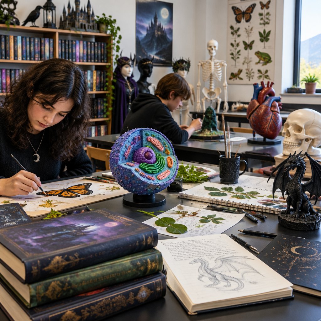
Biology art projects for visual learners turn abstract processes into something students can see, touch, and draw, and these biology art projects make the concepts stick. A 2026 study found that students who drew and modeled biological concepts outperformed peers taught through text alone (Art-Based Learning in Biology study). Here are seven projects, each with materials, time, and learning goals.
1. Clay Cell Model. Sculpt a plant or animal cell from clay, labeling organelles. Materials: clay, toothpicks, cardstock labels. Time: 45 minutes. Goal: cell parts and functions.
2. Organelle Watercolor Map. Give each organelle its own color and caption on a large cell diagram. Time: 30 minutes. Goal: memory through color coding.
3. Paper Skeleton Build. Cut and assemble a paper skeleton, then compare it to a real animal skeleton. Time: 50 minutes. Goal: anatomy and structure.
4. Habitat Diorama. A shoebox ecosystem showing animals, plants, and food webs. Time: 60 minutes. Goal: ecosystems and interdependence.
5. Plant Cell Collage. Torn paper and fabric scraps form cell walls, chloroplasts, and vacuoles. Time: 25 minutes. Goal: plant cell structure.
6. Animal Anatomy Sketchbook. Weekly observational drawings of bones, joints, and muscles. Time: 20 minutes per session. Goal: skeletal systems.
7. Life Cycle Scroll. A long paper scroll illustrating metamorphosis or plant growth. Time: 40 minutes. Goal: biological processes over time.
|
Project |
Materials |
Time |
Core Concept |
|---|---|---|---|
|
Clay cell model |
Clay, labels |
45 min |
Organelles |
|
Organelle watercolor |
Paints, paper |
30 min |
Cell functions |
|
Paper skeleton |
Cardstock, brads |
50 min |
Anatomy |
Time
For a fuller walkthrough, see how to teach biology using artistic models.
Visual Biology Learning Strategies That Make Concepts Stick
Visual biology learning strategies work because drawing forces retrieval, not just recognition. Research indexed by the National Center for Biotechnology Information links visual arts to stronger cognitive processing and memory. Three strategies we recommend:
- Draw-then-label: sketch first, add terms after.
- Model-and-explain: build, then teach a partner.
- Color-code systems: one color per organ or process.
Coloring books reinforce anatomy the same way; our guide on how anatomy coloring books help medical students learn explains the mechanism.
3D Printed Anatomical Models for Classrooms and Anatomy Teaching Tools for Educators
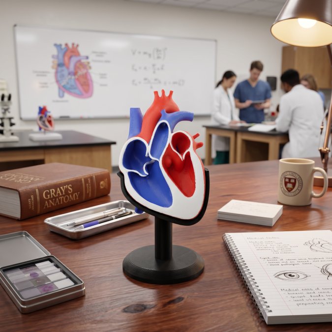
3D printed anatomical models for classrooms give students a physical structure to hold, rotate, and compare, which supports the visualization skills STEAM curricula now emphasize (Springer Nature STEAM study). When choosing anatomy teaching tools for educators, check three things:
- Durability under constant handling
- Accuracy of anatomical detail
- Fit with your grade level and lesson plan
LumaLayer Creations makes custom anatomical models for classrooms and clinics, making complex structures tangible. Start with our anatomy teaching tools guide for educators, or learn how to buy custom 3D printed anatomy models tailored to your lessons. If you want students to wear their learning, custom apparel printing is one of the services LumaLayer Creations offers.
Building a visual biology unit takes planning, but the payoff is students who remember what they built. LumaLayer Creations creates custom 3D printed anatomical models and original learning tools that make complex structures tangible for classrooms and clinics. Explore our custom printing and teaching models to bring hands-on anatomy into your next lesson. Get started with LumaLayer Creations and give every visual learner something to hold.
Frequently Asked Questions
What are some good biology art projects for visual learners?
Start with projects that turn abstract structures into something you can see and touch. Cell models made from clay or foam, labeled anatomy drawings, habitat dioramas, and plant life-cycle sculptures all work well. Each one asks students to translate a biology concept into a visual form, which strengthens comprehension and memory. Pair every project with a short written or verbal explanation so the art stays scientifically accurate.
How does art integration improve retention in biology?
Drawing and modeling force students to notice details they might skip when reading. Research on art-based learning suggests that drawing and physical modeling can improve learning outcomes compared with traditional methods. The act of building or sketching a cell or skeleton creates a visual memory anchor, which helps visual learners recall structures and functions long after the lesson ends.
Can 3D printed anatomical models support classroom art projects?
Yes. A 3D printed model gives students a durable reference they can hold, rotate, and sketch from multiple angles. Teachers use them as drawing prompts, comparison tools, and assessment aids. LumaLayer Creations makes custom 3D printed anatomical models for classrooms, which pair well with drawing and sculpture projects. Check durability and material safety before heavy student handling, and confirm pricing directly with the studio.

0 Comments