

Table of Contents
- Why Models Work: The Case for Tactile Learning in Anatomy
- Choosing the Right Models for Your Anatomy Lesson Plan with Models
- Anatomy Teaching Activities That Keep Students Engaged
- How to Run an Anatomy Model Labeling Activity
- How to Memorize Anatomy: Strategies That Stick
- Maintenance, Safety, and Making Models Last
- Differentiated Instruction and Budget-Friendly DIY Options
- Frequently Asked Questions
Last Updated: October 15, 2026
Why Models Work: The Case for Tactile Learning in Anatomy
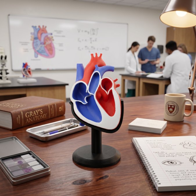
Tactile learning builds understanding through direct physical contact. Handling a physical model lets students map spatial relationships in a way flat diagrams cannot replicate.
Anatomy is a three-dimensional subject long taught with two-dimensional tools.
The research on this is straightforward.
A model is a tool, not a teaching strategy. Whether it transforms a lesson or gathers dust comes down to how the instructor builds activities around it.
Choosing the Right Models for Your Anatomy Lesson Plan with Models
Start with the learning objective, then work backward to the model. A small collection of focused models outperforms one large showpiece bought to serve every purpose.
Matching Model Types to Learning Objectives
Skeletal models work best for structure and landmark identification: bone names, joint types, and the axial-appendicular relationship. Muscular models suit movement, origin and insertion points, and muscle coordination. Organ system models (heart, lungs, digestive tract, brain) fit physiology lessons where function matters as much as form. A dissection alternative that comes apart in layers supports lessons on superficial versus deep structures.
Anatomy Teaching Activities That Keep Students Engaged
Engagement comes from structure, not novelty.
stack of three cat bowl risers
Anatomy teaching activities that consistently work include station rotations, peer teaching, timed identification challenges, and build-and-explain tasks. Each forces active recall rather than passive review.
Station Rotation and Group Work
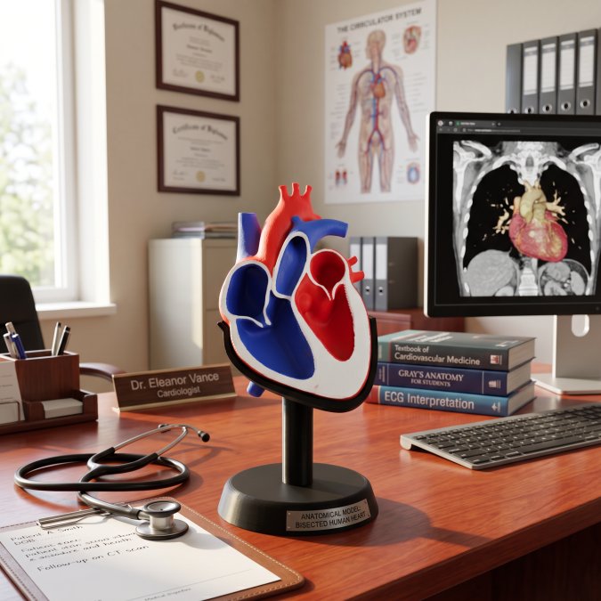
Set up four to six stations, each with a different model and a short task card, and rotate groups every eight to ten minutes. One station might ask students to identify five landmarks on a skeletal model; another to trace blood through a heart model; a third could use a digital-physical hybrid, where students check their identification against a 3D visualization on a tablet.
A few mechanics that make rotations run smoothly:
- Group size of three to four. Larger groups leave students watching; smaller groups leave stations idle.
- Assign roles inside each group. One student handles the model, one reads the task card, one records, one reports out. Rotate roles at each station so every student handles the model at least twice per class.
- Write task cards at three levels. Basic cards ask students to name structures; mid-level cards to describe relationships (“which structure sits deep to this one?”); advanced cards to predict a consequence (“what happens to this pathway if this vessel is blocked?”). Hand out the level that fits the group without announcing it.
Peer Teaching with Models
Peer teaching works when the explaining student has to physically point at structures while talking. Pair students and give each pair one model and one prompt: “Teach your partner the path of blood through the heart, using the model to show each step.” The listener holds a checklist of structures that must be named and pointed to.
Timed Identification Challenges
A timed challenge turns labeling into a game without chaos. Give each pair a model with labels removed and a numbered answer sheet, set a five-minute timer, and have students write the name for each numbered structure. When time is up, pairs swap sheets and score each other against your answer key. Structures missed across multiple pairs tell you exactly what to reteach, so tally the three most-missed and address them before the next activity.
Build-and-Explain Tasks
For lessons where function matters as much as form, have students build a simple structure from clay, foam, or paper and explain it to a partner. A student building a nephron from clay must decide where the loop of Henle goes and why, that decision-making is the learning. Pair the build with a short written explanation so you have something to assess beyond the model itself.
Choosing the Right Activity for the Objective
Match the activity to the verb in your learning objective. “Identify” fits a timed labeling challenge; “explain” fits peer teaching or build-and-explain; “compare” fits a station rotation with two related models at one station.
How to Run an Anatomy Model Labeling Activity
An anatomy model labeling activity is the most efficient way to check whether students can actually identify structures, not just recognize them. Use removable labels, sticky flags, or numbered pins rather than permanent markers.
- Prepare models with all labels removed
- Number each structure position with a pin or flag
- Distribute answer sheets with matching numbers
The real value is in the correction step. When a student mislabels the radius and ulna, you can address the confusion immediately, while the model is still in their hands.
How to Memorize Anatomy: Strategies That Stick
Knowing how to memorize anatomy is a skill separate from understanding it, and students need both. The most effective strategies combine the model with active recall and spaced repetition. Clinical correlation is the strongest memory anchor: tie the brachial plexus to a common injury pattern and each cranial nerve to a function students can test on themselves. Physiological processes give structure to memorization, have students trace digestion or circulation through a model in sequence, and the sequence itself becomes the memory framework.
Spaced review matters too: a five-minute labeling sprint at the start of each class, using a different model each time, does more for long-term retention than a single cram session.
Maintenance, Safety, and Making Models Last
Durability is where most buying guides go quiet. Models handled by dozens of students every week will wear, and how you clean, store, and repair them determines whether they last one semester or several years.
Cleaning by Material
Different materials need different cleaning methods; the wrong one is the fastest way to ruin a model.
- Hard plastic and resin models (skeletal, organ): Wipe with a soft cloth dampened with mild dish soap and warm water. Avoid alcohol, acetone, or abrasives, which cloud surfaces and dissolve painted labels. Use a soft-bristle toothbrush for crevices.
- Painted or hand-finished models: Damp-wipe only; never submerge. Paint can lift if soaked, and once a label or color code is gone, the model loses teaching value.
- 3D-printed models (PLA, PETG, resin): PLA softens at low temperatures, so keep these out of hot cars, direct sun, and dishwashers. Wipe with a damp cloth. Resin prints are more brittle than filament prints and need extra care around thin features.
Wipe down models at the end of each lab day, deep clean at the end of each unit, and inspect removable parts monthly.
Safety Protocols for Handling Models
Safety protocols for handling models are simple but non-negotiable. Wash hands before and after handling, keep models away from food and drink, and inspect removable parts regularly for cracks or loose connections, especially small detachable pieces that can become a choking hazard in younger classrooms.
- Small parts: Inventory any detachable piece smaller than a golf ball before and after each lesson, and sweep for missing pieces before students leave.
- Shared handling: Wipe models between groups to reduce germ spread; keep a spray bottle of mild soap solution and microfiber cloths at the front of the room.
- Sharp or pointed features: Some skeletal and dissection-alternative models have wire pins, hinges, or pointed processes. Demonstrate safe handling before the first activity and set a clear rule: no pointing a model feature at another student.
Storage That Actually Protects Models
Storage is the biggest factor in longevity. Store models in closed cabinets or covered bins away from direct sunlight, which degrades many plastics. Keep small parts in labeled bags inside the main container, and never force moving joints past their natural range, the most common cause of breakage.
- Store by system, not by size. All skeletal models together, all organ models together, so it’s faster to pull a set and easier to spot a missing piece.
- Use clear bins with labels on the short end so you can read them without pulling the bin off the shelf.
- Keep a desiccant packet in bins holding foam or fabric-lined models. Moisture is the enemy of long-term storage.
Simple Repairs You Can Do Yourself
Most classroom model damage falls into three categories, all with straightforward fixes.
- Loose or detached small parts: A small dot of cyanoacrylate (super glue) or two-part epoxy reattaches most plastic parts. Clean both surfaces, apply a thin layer, hold for thirty seconds, and let cure overnight before returning the model to student use.
- Cracked or split pieces: For non-structural cracks, a thin bead of epoxy along the crack line, sanded smooth after curing, restores the surface. For structural cracks in a load-bearing area, retire the model from student handling and use it for demonstration only.
- Worn or faded labels: A fine-tip paint pen in a matching color can restore printed labels. Keep paint pens in the manufacturer’s colors (typically red for arteries, blue for veins, yellow for nerves) so touch-ups blend in.
Knowing When to Retire a Model
Not every model is worth saving. Retire a model when a repair would cost more time than it’s worth, when a structural failure makes it unsafe for student handling, or when missing parts make the intended objective impossible to teach. A model with three of ten removable organs left is clutter, not a teaching tool. Move it to a demonstration-only shelf.
A Maintenance Calendar That Fits a School Year
A simple rhythm keeps a classroom set in working order without adding much to your week:
- Daily: Quick wipe-down after each lab, small-parts count before dismissal.
- Weekly: Inspect removable parts for cracks or looseness, and restock missing pieces from your spare-parts bin.
- End of unit: Deep clean each model used, photograph the complete part set, and update bin labels.
That calendar is the difference between a model set that lasts one year and one that lasts five.
Differentiated Instruction and Budget-Friendly DIY Options
Not every classroom has a full set of commercial models, and not every student learns the same way.
For differentiated instruction, assign the same model but vary the task.
This is also where custom 3D-printed anatomical models earn their place.
LumaLayer Creations builds custom 3D-printed anatomical models designed for real classroom handling, alongside original designs that blend science and craft.
Frequently Asked Questions
What are some fun ways to teach anatomy and physiology with models?
Turn model work into a game. Try timed assembly races where teams rebuild a disassembled skeletal model, or a ‘mystery structure’ challenge where students identify a covered part by feel. You can also run a clinical correlation scenario: give students a patient symptom and have them point to the affected organ system on the model. These anatomy teaching activities boost engagement and retention far better than lecture alone.
How do you teach students to identify structures on an anatomy model?
Start with a guided tour where you name and point to each structure while students follow along on their own models. Then move to a labeling activity: provide color-coded stickers or removable labels and have students place them correctly without notes. Finish with a peer quiz where one student points and the other names the structure. This progression from guided to independent practice builds durable recall.
What is the best way to memorize anatomy for exams?
Combine spatial and verbal memory. Handle a physical model while saying the structure name aloud, then cover the labels and test yourself. Spaced repetition helps too: review the same model for 10 minutes daily rather than cramming once a week. Students who use models alongside flashcards consistently outperform those who rely on either method alone.
How can teachers use anatomical models in a lesson plan?
Begin with a short lecture introducing the system, then transition to hands-on model exploration in small groups. Assign each group a different body system and have them present their model to the class. Wrap up with a formative assessment where students label a blank diagram or answer questions using only the model as reference. This structure works for a single class period or a multi-day unit.
Are 3D-printed anatomical models durable enough for classroom use?
Yes, when printed with appropriate materials and handled with care. PLA and PETG prints hold up well under normal classroom conditions. Store models in padded containers, avoid leaving them in hot vehicles, and clean with a damp cloth rather than harsh solvents. With basic maintenance, a well-made 3D-printed model can last through multiple semesters of regular student handling.
What activities help students learn anatomy with 3D models?
Try dissection alternatives where students explore layers of a 3D-printed organ model instead of a specimen. Model assembly challenges teach spatial relationships between structures. Digital-physical hybrid activities pair a physical model with a tablet app so students can cross-reference. Each of these anatomy teaching activities targets a different skill: identification, spatial reasoning, and clinical application.
0 Comments