


Table of Contents
- Why 3D-Printed Anatomical Models Help Medical Students Visualize Complex Structures
- How to Choose the Best Anatomical Models for Your Study Goals
- 3D-Printed Heart Model for Medical Education: Tracing Vessels and Chambers
- 3D-Printed Brain Anatomy Model: Mapping Lobes, Vessels, and Deep Structures
- 3D-Printed Human Body Model for Anatomy Study: Whole-System Learning
- Ranked Comparison: Which Model Types Suit Which Students
- Cost and Access: 3D-Printed Models vs. Atlases, Digital Tools, and Cadavers
- What the Evidence Says About Learning Outcomes and Limitations
- Frequently Asked Questions
Last Updated: October 17, 2026
Why 3D-Printed Anatomical Models Help Medical Students Visualize Complex Structures
Anatomical models are physical representations of the body’s structures, built to scale so students can hold, rotate, and disassemble them. At LumaLayer Creations, we build custom 3D-printed anatomical models for educators who need students to see what a flat page cannot show.
3d printed gothic style pendant featuring cat skulls and flowers
The core problem is spatial. A textbook drawing of the brachial plexus flattens a three-dimensional tangle of nerves into a single plane. A printed model does not.
Below, we break down the model types that work best for study, how to choose between them, and where printed models fit against atlases, digital tools, and cadavers.
How to Choose the Best Anatomical Models for Your Study Goals
Start with the exam or course objective, then work backward to the model. A first-year student memorizing bony landmarks needs something different from a surgical resident rehearsing an approach.
Anatomy Level, Scale, and Color Coding
Match the model to the depth of your syllabus. Introductory courses benefit from simplified, enlarged models where key structures are exaggerated. Advanced study needs finer detail and correct proportions.
- Scale: Enlarged models (2x to 4x) help beginners isolate small structures. Life-size models suit exam rehearsal and clinical reference.
- Color coding: Distinct colors for arteries, veins, and nerves speed recognition. Some students prefer single-color models so they must identify structures without color cues, which better mimics exam conditions.
- Disassembly: Removable parts let you quiz yourself. A model that comes apart into labeled pieces supports active recall far better than a solid cast.
Materials, Resolution, and Durability
Most printed teaching models use various filaments. Some are rigid and print with sharp detail but may soften in heat. Others handle classroom handling and minor drops better. Some printing methods deliver finer surface detail for small structures, though parts may be more brittle.
Resolution matters most for tiny features. A finer layer height captures thin vessel walls and delicate nerve branches. Durability matters most for shared classroom sets, where models pass through many hands each semester. Ask how a model was post-processed, since sanding and coating affect both feel and longevity.
3D-Printed Heart Model for Medical Education: Tracing Vessels and Chambers
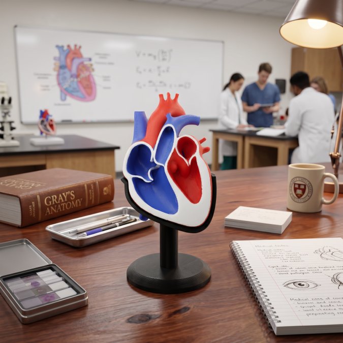
A 3D-printed heart model for medical education is the single most useful printed study aid for cardiovascular anatomy, because the heart rewards spatial reasoning more than almost any other organ.
The best heart models split into halves or quarter sections. That lets you open the organ, follow blood from the right atrium through the tricuspid valve into the ventricle, then out the pulmonary trunk.
Look for models that show the aortic arch and its branches clearly, since exam questions often hinge on vessel relationships. A removable anterior wall is worth more than extra surface detail.
3D-Printed Brain Anatomy Model: Mapping Lobes, Vessels, and Deep Structures
A 3D-printed brain anatomy model earns its place when it separates into lobes and exposes deep structures that diagrams stack confusingly. The cortex is easy to sketch. The limbic system, ventricles, and internal capsule are not.
Choose a model that breaks into hemispheres and includes a removable brainstem and cerebellum. Deep structures like the basal ganglia and thalamus should be color-differentiated so you can name them in place.
What most guides miss is how much a physical brain model helps with orientation. Rotating a printed hemisphere in your hands builds the mental rotation skill that imaging interpretation demands.
3D-Printed Human Body Model for Anatomy Study: Whole-System Learning
A 3D-printed human body model for anatomy study works best as a systems overview rather than a detail tool. Torso models with removable organs let students see how the respiratory, digestive, and cardiovascular systems stack and share space.
These models suit survey courses and exam review. Pull the lungs, lift the liver, and the spatial relationships between organs finally click. Smaller desktop torsos fit dorm study, while larger classroom versions support group quizzing.
Pair a whole-body model with focused organ models. The torso gives context; the heart and brain models give depth. Together they cover gross anatomy from overview to close study.
Ranked Comparison: Which Model Types Suit Which Students
Here is how the main model types stack up for different study needs.
|
Model Type |
Best For |
Key Strength |
Watch Out For |
|---|---|---|---|
|
Heart model |
Cardiovascular units, exam prep |
Vessel and chamber relationships |
Choose one that splits open |
|
Brain model |
Neuroanatomy, imaging courses |
Deep structure orientation |
Confirm deep structures are shown |
|
Whole-body torso |
Survey courses, review |
System relationships |
Weak on internal detail |
|
Skeletal model |
Bony landmarks, joints |
Palpation and landmark practice |
Limited soft tissue |
|
Custom patient-specific model |
Case study, surgical planning |
Real anatomical variation |
Needs imaging data to build |
Ranked by everyday study value for most medical students: heart first, brain second, skeletal third, whole-body torso fourth, custom models for specific cases. Your syllabus may reorder that list, so check your exam objectives before buying.
Cost and Access: 3D-Printed Models vs. Atlases, Digital Tools, and Cadavers
Printed models sit in the middle of the cost and access spectrum, and the trade-offs are more concrete than most guides admit. Here is how the four main study resources compare on what a student actually spends and what access they require.
|
Resource |
Typical Upfront Cost |
Ongoing Cost |
Access Requirement |
Best Use |
|---|---|---|---|---|
|
Printed anatomical model |
Varies by size and material; a single desktop model is the lowest-cost physical option |
Near zero after purchase |
None beyond a desk |
Repeated self-testing, spatial rehearsal |
|
Printed atlas |
Low, one-time |
Replacement if damaged |
None |
Labeling reference, exam review |
|
Digital anatomy app |
Low subscription or one-time app fee |
Subscription renewals |
Device and internet |
Quizzes, cross-sections, labeling drills |
|
Cadaver lab |
Included in tuition at most programs |
None to the student directly |
Enrollment, lab scheduling, supervision |
Tissue realism, variation, dissection technique |
The pattern most students land on: atlases and apps cost least and travel easily, cadavers offer unmatched realism but are gated by lab access and scheduling, and printed models give you a physical object you can handle any time for a one-time outlay. A printed model does not need a lab slot, a supervisor, or a charged battery.
Where the Money Actually Goes
For a printed model, the cost drivers are size, complexity, material, and whether the design is stock or custom. A small single-organ model can be a less expensive path. A multi-part torso or a patient-specific model built from imaging data may cost more because of design and print time.
Maintenance can be a consideration. Shared classroom sets may take handling damage, so budgeting for occasional replacement of small removable parts can be wise. Models made with certain materials and finishes may outlast others in a shared lab setting, which can affect the real cost over time.
Access Angles Students Overlook
- Library and lab lending: Some anatomy departments and health-sciences libraries lend printed models the way they lend reserve textbooks. Ask before buying.
- Makerspaces: Campus makerspaces and public library fabrication labs sometimes print educational models for a materials fee, which can undercut buying a finished model.
- Group buys: Splitting a multi-part model set across a study group lowers per-student cost and gives each person a piece to quiz from.
- Digital-first, print-second: Buy or borrow the digital file first, then print only the structures you keep mislabeling. This avoids paying for detail you already know.
For schools buying for multiple systems, ask about educational and bulk arrangements directly rather than assuming list pricing applies.
What the Evidence Says About Learning Outcomes and Limitations
The evidence for printed models is encouraging but not conclusive, and the useful question is not whether they work but where they work. Educators consistently report that hands-on models improve engagement and spatial understanding, and many anatomy programs now use printed models alongside traditional resources. Formal studies on learning outcomes vary in size and design, so treat strong claims with caution and look at the mechanism instead.
Why Physical Models Help (The Mechanism)
Spatial anatomy is a mental rotation problem. A textbook drawing fixes one viewpoint; a printed model lets you rotate the object and rebuild the relationship from any angle. That practice transfers to imaging interpretation, where students must mentally slice and reorient structures. The tactile loop also supports active recall: tracing a vessel with a fingertip and then naming it without a label is a self-test, not a reread.
Where Printed Models Fall Short
- Simplification: Printed models simplify real anatomy. A clean model of the brachial plexus omits the small branches and connective tissue that make dissection hard.
- Limited variation: A single model represents one body. Real anatomy varies by age, sex, body type, and individual variation, and a model that never varies can teach a false sense of uniformity.
- No tissue realism: Printed plastic cannot replicate the texture, friability, and plane-cleavage of real tissue, which is why dissection remains the standard for surgical technique.
- Label dependence: Models that come pre-labeled can encourage recognition rather than recall. Test yourself with labels covered or removed.
How to Test Understanding Against Real Variation
Use printed models to build the spatial map, then stress-test it. Compare the model against atlas plates of variant anatomy, against imaging cases that show the same structure from a different angle, and against any cadaver or prosection access your program offers. If you can describe a structure from memory, rotate it mentally, and predict how it would look on a scan, the model did its job. If you can only name the labeled parts, you memorized a model, not anatomy.
Frequently Asked Questions
What are the most useful 3D-printed anatomical models for medical students?
The most useful sets combine a heart model for tracing chambers and blood vessels, a brain model for mapping lobes and deep structures, and a full-body or regional model for whole-system review. Students preparing for gross anatomy exams benefit most from models that disassemble into layers, because taking a structure apart and rebuilding it builds spatial understanding faster than reading a labeled atlas page. Start with the system you find hardest to picture, then add models as your coursework moves through each region.
How do 3D-printed anatomy models help students understand complex structures?
Printed models turn a flat diagram into an object you can rotate, hold, and take apart. That tactile learning reinforces spatial understanding of how bones, organs, and blood vessels sit in relation to one another, which is difficult to build from a textbook alone. Color-coded and disassemblable models let you isolate one structure at a time, then reassemble the whole system. Many students use them alongside cadaver study or digital tools rather than as a replacement, treating the model as a rehearsal before lab sessions.
What features should students look for in an anatomy model?
Check four things: anatomy level (does it match your course, from undergraduate survey to medical gross anatomy), scale (life-size versus enlarged for fine detail), color coding (does it distinguish arteries, veins, and nerves clearly), and disassembly (can you remove layers to see internal structures). Also weigh material and resolution. A higher-resolution print may show finer vascular detail but could be more fragile, while a tougher material might survive constant handling in a study group. Consider how the model is finished, as this can affect both durability and how clearly small features read.
How do 3D-printed anatomical models compare with traditional anatomy teaching aids?
Traditional plastic models are mass-produced and consistent, but often limited to standard systems and fixed configurations. 3D-printed models offer more flexibility: patient-specific or custom structures, unusual variations between bodies, and replacement parts when something breaks. Cost and access vary widely depending on material, size, and whether you print in-house or order a finished model, so compare against atlases and digital tools for your budget. The strongest approach is usually mixed: printed models for hands-on spatial practice, atlases for labeling, and digital tools for quizzes.
0 Comments